Session 1: Andrew Stevenson (University of Western Australia)
New Approaches for Regenerative Medicine; chaired by Lynn Wise and Chris Turner
Session 1 began with a plenary from Phil Stephens from Cardiff University, who spoke about the unique healing properties of the oral mucosal cavity. In particular, he informed us of the superior wound healing properties of a small subset of oral fibroblasts, called oral progenitor cells, and their potential benefit to wound healing and tissue repair therapies. He was followed up by Alison Cowin from the University of South Australia, who spoke to us about some of the work she was doing with the Wound management CRC on stem cell therapy. Michael Samuel form the Centre for Cancer Biology spoke next, and he informed us about the work his group has been doing on the Rho-ROCK pathway in wound healing, working with an inhibitor of ROCK signalling, 14-3-3ζ, which has shown promising results increasing wound healing in normal and diabetic wounds. Andrew Stevenson was the next speaker, and he spoke about the use of a lysyl oxidase inhibitor (LOXi) in the treatment of fibrosis and scarring. Lysyl oxidases (LOXs), catalyse the cross-linking of collagen and his group has been using a LOXi to inhibit this process in order to improve scar function and appearance, which has has shown promise in in vitro, as well as in mouse and pig models. The final speaker of the session was Ilia Banakh, who gave an excellent talk on the comparison of many commercially available dermal substitutes such as Integra and Novosorb BTM, and look at wound contraction, vascularisation, host cell infiltration, localised growth factors concentrations and ECM deposition.
Session 2: Carla Canizzo (University of Sydney)
Cutaneous Melanoma; chaired by Mitchell Stark and Joseph Rothnagel
Following afternoon tea of fresh fruit and freshly baked muffins we attended session 2 on cutaneous melanoma chaired by Mitchell Stark and Joseph Rothnagel. Plenary speaker Professor Claudia Wellbrock from the University of Manchester highlighted the need to understand intra-tumour signalling between melanoma cells and non-tumour cells as it has a major impact on MAPK-pathway therapies. Professor Wellbrock and her group have found that the melanoma specific transcription factor microphthalmia-associated transcription factor (MITF) can cause MAPK-pathway therapy resistance and can therefore be used to classify melanoma-phenotypes as ‘EMT like’ or non ‘EMT like’. She also highlighted that the heterogeneity of these phenotypes can also lead to resistance to BRAF inhibitors, therefore accelerating tumour growth and metastases. In addition, Dr. Loredana Spoerri from the Diamantina Institute explained that high MITF levels display a homogenous distribution of cell cycling while low levels of MITF are characterised by clusters of cycling cells and G1-arrested cells. Dr. Spoerri explained that MITF over-expression gives rise to flatter spheroid structures in vitro, however this did not reduce the inner hypoxic levels and therefore G1 arrest. Hence, high MITF levels prevent cell cycle arrest due to the hypothesised cell-cell and extr-cellular matrix cross-talk.
Furthermore, Dr. Imanol Arozarena from Navaarabiomed spoke about the complex nature between tumour cell metabolism and melanoma progression. He highlighted that many studies have explored the link between high glucose and melanoma risk however the link between glucose depletion on specific proteins or signalling proteins remained unknown. He focused on his groups most recent findings that glucose restriction can induce a “MITFlow/AXLhigh” phenotype that is more slow and aggressive and more likely to be therapy resistant.
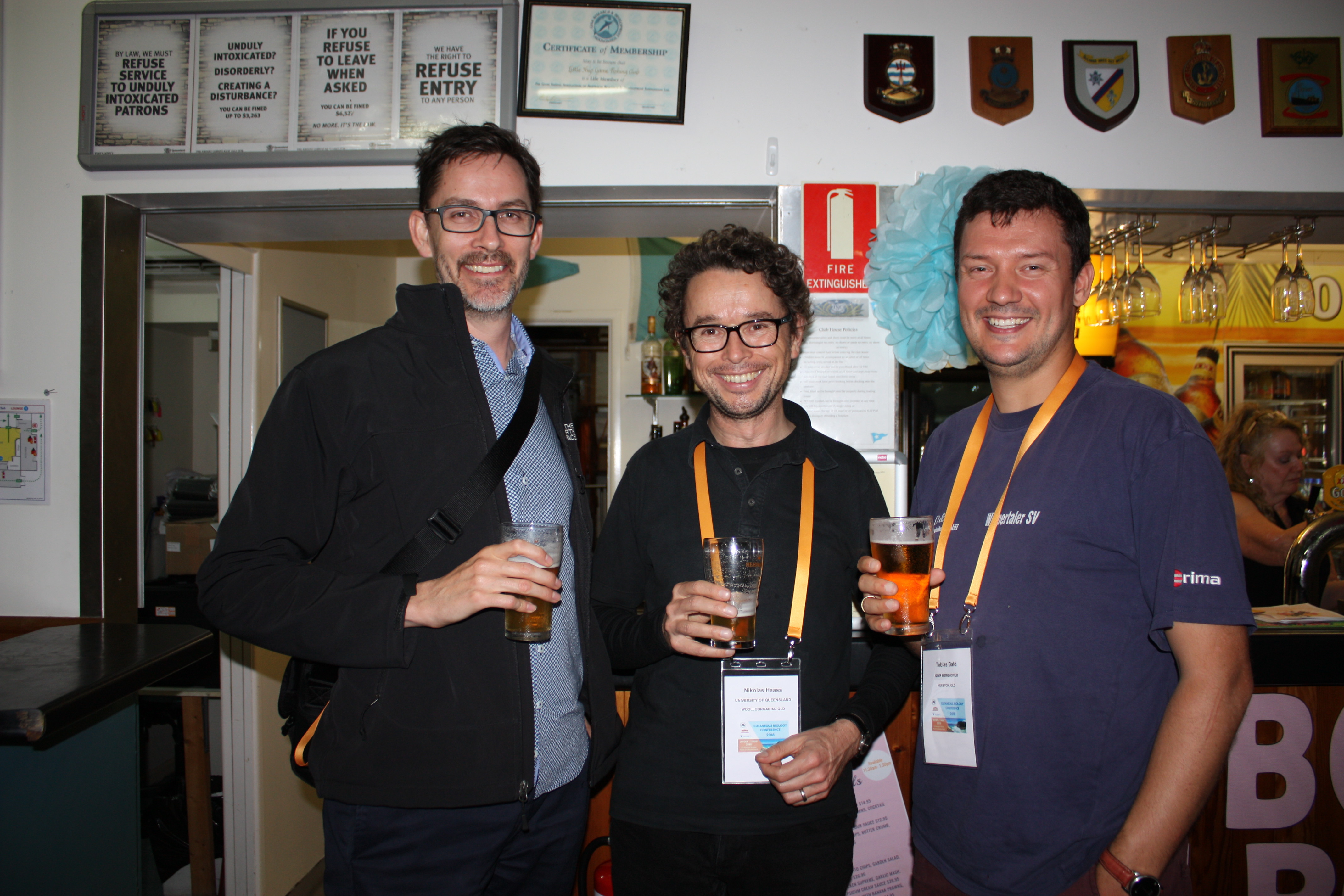
On the topic of cancer therapy resistance, Professor Nikolas Haass from the Diamantina Institute explained that advanced stage melanoma has poor prognosis and that there is a need to identify new therapeutic strategies. His group has found that the GTPase RAB27A is overexpressed in melanomas and this correlates with poor patient survival. His studies further highlighted that loss of RAB27A expression in melanoma cell lines inhibits 3D spheroid invasion and cell motility and metastasis both in vitro and in vivo respectively. More interestingly however, is that RAB27A is expressed higher in melanoma than other cancers and therefore these findings suggest that RAB27A may be used as a biomarker or therapeutic marker for melanoma.
Continuing on, Sheena Daignault also from the Diamantina Institute spoke about enforcing cellular stress to promote apoptotic and immunogenic responses in melanoma. Her studies involved promoting ER stress and apoptosis with Bortezomib in melanoma to induce immunogenic cell death. In her experiments, mice were primed with Bortezomib-treated melanoma cells and then following one week with live melanoma cells. Sheena highlighted that primed mice had a delay in tumor growth and volume when compared to controls, suggesting that immunogenic cell death may be used as a therapeutic method for treating melanoma.
Dr. Dubravka Skalamera from the Mater Medical Research Institute explained how her established microscopy platform has allowed for high resolution images of melanocyte, keratinocytes and fibroblasts. More interestingly however is that this technique is able to analyse the effects of melanoma-associated mutations on the phenotype of cells. For example her group found that HACAT keratinocytes and wild type melanocytes form dendrites for molecular exchange, however this phenotype is reduced with the expression of NRAS.Q61K in melanocytes. Furthermore dermal fibroblasts reduced melanocyte proliferation and increased apoptosis, however the NRAS.Q61K expressing melanocytes did not show any of these characteristics highlighting its role in cancer progression.

Session 3: Dr Chris Turner (University of British Columbia)
Wound Repair; chaired by Allison Cowin and Anjana Bairagi
It was an absolute pleasure to be a participant at Cutaneous Biology 2018 and I’m grateful to the AWTRS for the Travel Award. As an ex pat Aussie now living in Canada, I was afforded the opportunity to see former colleagues, old friends and revisit the joy of meat pies, tinned beetroot and fairy bread.
Session 3 showcased a great selection of speakers. The plenary, Kiarash Khosrotehrani (University of Queensland), started us off by delving into the genetic, cellular and environmental determinants of wound closure, reinforcing the complexity of wound healing and in particular microbial colonization and scarring. Next up, Mark Fear (Fiona Wood Foundation) took us on an entertaining and often comedic journey into fibroblasts, teasing apart the specific subpopulations and how these affect fibrotic responses. Leila Cuttle (Queensland University of Technology) spoke of LC-MS/MS in conjunction with a host of other state of the art techniques to investigate gene expression and key analytes in wound fluid. I was particularly interested in her wound fluid biobank, a very valuable resource. Stuart Mills (University of South Australia) nicely introduced the importance of inflammation as an important factor in mediating transition of wounds to a chronic phenotype. The actin remodelling protein, Flightless, was uncovered as having an interesting a role in macrophage polarization, one with potentially important consequences for chronic wound repair. To end the session, Ruilong Zhao (Sutton), confronted us with very graphic images of burn survivors, reminding us how important our research into all areas of cutaneous biology really is. Here we learnt about Protein C, and how plasma expression may be a better disease course predictor than burn size, depth and level of inhalation injury alone.
I also had the pleasure of attending the EMCR Workshop led by Hugh Kearns from iThinkWell about ‘Developing a research track record on a shoestring’. Sponsored by the Australian Academy of Science Theo Murphy Initiative, this educational and highly motivational session made me want to get back into the lab and finish off those last experiments needed for that unfinished manuscript. We learnt about goal setting and importantly for me prioritization of work.
Session 4: Dr Natalie Stevens (University of South Australia)
Tissue Engineering; chaired by Brooke Farrugia and Aleta Pupovac
Following Morning Tea, an interesting and varied session on Tissue Engineering was held where five female researchers showed exciting technological advances in wound care and regeneration. The session’s plenary speaker Magda Ulrich from the Association of Dutch Burn Centres in Amsterdam excellently summarised the need for dermal substitutes as alternatives to skin grafts in the context of burn wounds. Her work assessing the use of epidermal and mesenchymal stem cells as part of dermal substitutes shows promise for the development of personalised fit-for-purpose dermal matrices which can reduce scarring and improve healing time and helps to elucidate the contributions of these stem cell populations in regeneration. Aleta Pupovac from CSIRO gave an interesting talk summarising an ambitious and innovative approach towards the development of skin models that incorporate microfluidic platforms for sampling and drug delivery, real-time imaging and innate immune components. This exciting technology could be the answer to reducing the use of animals in wound studies, which many AWTRS members would gladly welcome! Next, student Jiah Shin Chin from Nanyang Technological University in Singapore described her research using polycaprolactone (PCL) and rat collagen scaffolds, formed by elegant-looking wet electrospinning. Interestingly, the use of rat collagen with PCL reduced re-epithelialisation and prevented the formation of blood vessels and ECM through the scaffolds. Our incoming AWTRS president, Brooke Farrugia from the University of NSW, showed us the value of persistence and incidental findings with her work on chondroitin sulphate (CS) proteoglycans, which have roles in ECM remodelling. Her finding that mast cells express unique CS epitopes lead to the discovery of the role of the HYAL4 enzyme in mast cells which digests CS proteoglycans and provides evidence for a direct role of mast cells in collagen remodelling. The session was closed with a talk by Xanthe Strudwick from the UniSA Future Industries Institute who presented her interdisciplinary work on developing plasma coated dressings for wound healing. Her in vitro experiments characterised the differential effects of different coatings on fibroblasts and keratinocytes in culture and in vivo studies showed direct impacts on wound healing, indicating the different plasma coatings could be used to tailor wound dressings for different responses.
Session 5: Dr Stuart Mills (University of South Australia)
Photobiology and Skin Cancer; chaired by Vivienne Reeve and Shelley Gorman
Session 5 began with an international plenary speaker, Chikako Nishigori, from Kobe University in Japan, who was sponsored by RSC Publishing. Chikako gave a fascinating talk on the mechanisms of photocarcinogenesis and sunburn resolution. Her group studied skin cancers caused by UVB radiation and found that there were no specific UV induced mutations but that there was 8-oxoguanine formation and increased expression of the inflammatory markers 3-nitor-L-tyrosine. Using Ogg1 KO mice and UVB irradiation they discovered there was increased risk of skin carcinogenesis, associated with p53 mutations, and an increased expression of IL-1β and IL-6. Following this presentation James Wells, from the University of Queensland Diamantina Institute, talked about the mechanisms of CD8 T-cell mediated regression in squamous cell carcinomas (SCC) in a transplant model. They found that mice treated with Tacrolimus, an immunosuppressant given to transplant patients, where more susceptible to SCC formation. Interestingly, when the Tacrolimus treatment was withdrawn in the mice the tumours were rejected. Next to speak was Katie Dixon, from the University of Sydney, who had looked at targeting melanoma metastasis with vitamin D. Interestingly, she had discovered that people with higher sun exposure had higher vitamin D levels, as might be expected, but also that they had a better chance of tumour survival. The increase in the active form of vitamin D, (1,25D) in these people also had a concurrent increase in PTEN levels, which inhibited pAKT expression and blocked melanoma growth. This suggests that limited sun exposure to increase vitamin D levels and increase the protection against melanoma formation is more beneficial than little or no sun exposure. Following on from this Edwige Roy, from the University of Queensland, spoke about the epidermal clonal size after UVB exposure. He had found that the most proliferative cells after UV exposure were found around the hair follicles, which had larger clones and faster cycling cells and that this did not affect mutation burden but did initiate hedgehog induced carcinogenesis. Shelley Gorman, from the Telethon Kids Institute, was the next to speak on harnessing low dose UVB irradiation to curb obesity and metabolic dysfunction healing. UVB has been shown to reduce cholesterol inhibit diabetes and what Shelley was able to show was this this was vitamin D independent but instead was dependent on nitric oxide (NO) synthesis. This was confirmed in studies where she used a NO scavenger and weight loss in the mice was inhibited. The final speaker of the session was Felix Marsh-Wakefield from the University of Sydney. He had investigated how phototherapy altered circulating B cells to prevent clinically isolated syndrome (CIS) patients developing multiple sclerosis (MS). He showed, using mass spectrometry that there were 4 subsets of B cells, which were potentially altered by phototherapy which may be responsible for the delay in the onset of MS in CIS patients.

Session 6: Xanthe Strudwick (University of South Australia)
Stem Cell Biology; chaired by Kiarash Khosrotehrani and Stuart Mills
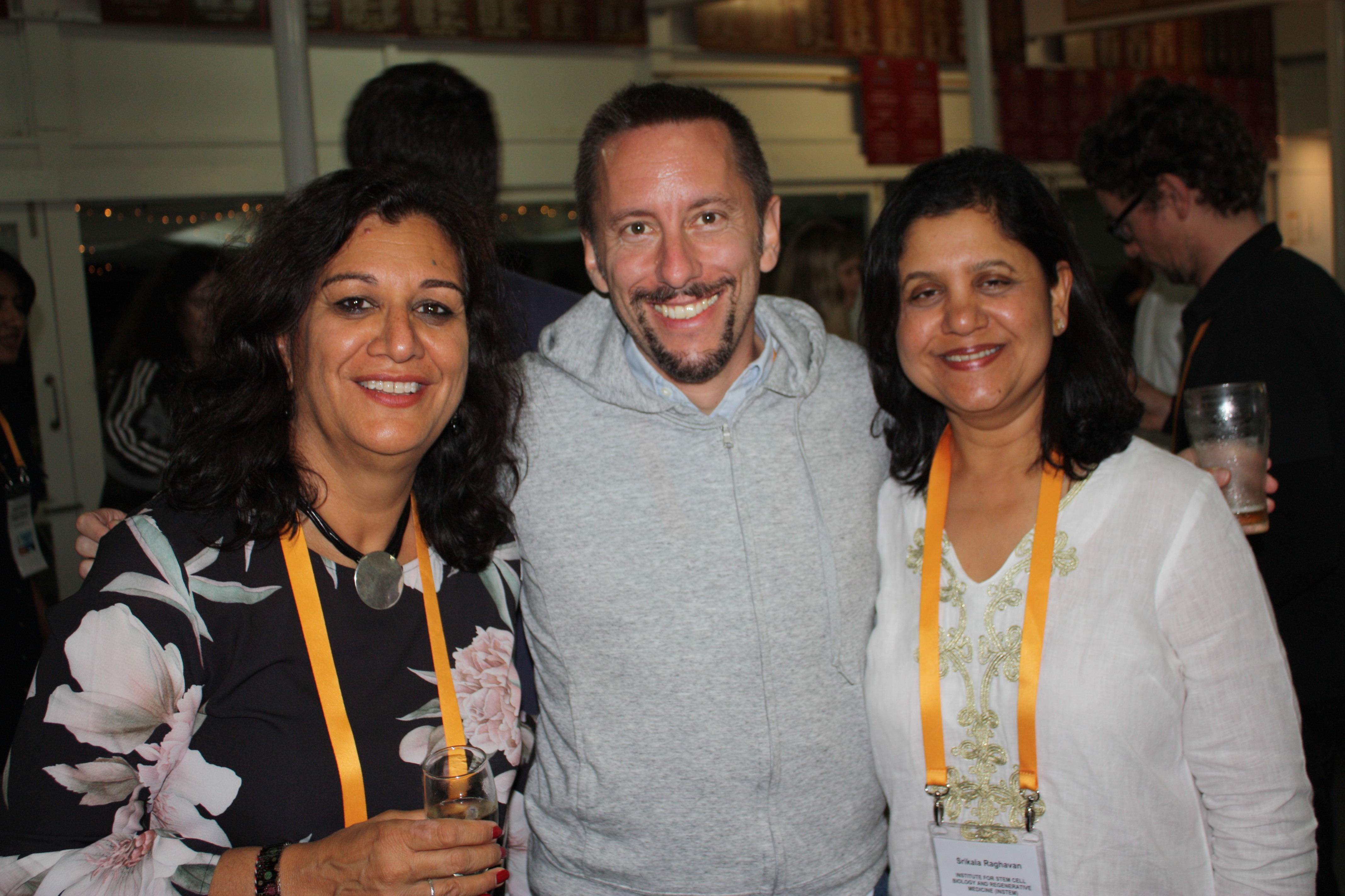
Wednesday morning began with Session 6 on Cell Biology which was chaired by Kiarash Khosrotehrani and Stuart Mills. The opening Plenary from Kim Jensen from the University of Copenhagen, Denmark, entitled ‘tracking stem cell during epidermal morphogenesis and homeostasis’, described the hair follicles’ role in the maintenance of functional epithelium from development, throughout life. He revealed how each area of the hair follicle is maintained as independent functional units by discrete in situ stem cells, but that this tissue compartmentalisation is reversible, such that it can be broken by injury, allowing the cells to contribute to the wound healing response. Moreover, each functional unit is maintained with its own homeostatic pace of division, which can be disrupted when a mutation in the K-Ras gene causes more proliferative and less differentiated cells to be produced. Pritinder Kaur from Curtin University then spoke about her research which shows that the ‘decreased tissue regenerative potential of ageing human skin can be attributed to changes in the dermal microenvironment’. She showed that the ageing phenotype is associated with the loss of pericyte numbers from the dermis as well as a decrease in pericyte expression of Versican and Lumican. Interestingly, this cell type which is usually associated with angiogenesis, can promote better basement membrane formation by keratinocytes and can promote epidermal formation when utilised as the sole ‘feeder’ cell in 3D culture due to their production of Versican. Srikala Raghavan then presented her research into the ‘role of Vinculin in regulating bulge stem cell quiescence’, where it was shown that the hair cycle is faster in Vinculin knockout mice, due to reduced telogen phase, a loss of quiescence t stem cells and subsequently increased proliferation. As Vinculin facilitates cell-cell adherens junctions, it appears that mutations in the Vinculin gene lead to a nuclear localisation of YAP and reduced alpha catenin recruitment to cytoskeleton, resulting in decreased cellular adhesions and lower numbers of quiescent stem cell in the hair follicle bulge. Finally, to round out the morning’s session, Zalitha Pieterse from Pritinder Kaur’s laboratory at Curtin University spoke about her project investigating the potential of human dermal pericytes as a feeder layer for ex-vivo expansion of patient keratinocytes. Under the alternative title of ‘Struggles of a 1st year PhD student’ she described the approaches taken to optimise the culture conditions and techniques to generate senescent feeder pericytes to use in the model system, before moving on to investigating the potential to replace 3T3 feeder layers with human pericytes for ex vivo patient keratinocyte expansion prior to autologous transplantation.
Session 7: Zalitha Pieterse (Curtin University)
Frontiers in Cutaneous Biology I; chaired by Nikolas Haaus and Lisa Tom
Session 7 of the Cutaneous Biology 2018 Conference Meeting was held on Wednesday morning 31st October 2018. This session was chaired by Nikolas Haas and Lisa Tom, hosted excellent talks around the theme of Frontiers in Cutaneous Biology. The session commenced with our Plenary Speaker, Stuart Pitson from the University of South Australia sharing about his lab’s work on targeting sphingolipids as a means to accelerate wound healing. Following on from this were two fantastic talks by Helmut Schaider and Mitchell Stark from the University of Queensland. Helmut introduced his work on how epigenetic remodelling leads to MAPKi-resistance in melanoma, specifically how they identified two potential candidates, OGT and TET1, for targeting drug resistance in melanoma. Mitchell Stark shared an exciting platform known as whole-exome sequencing and how this technology is used to identify the underlying genetic mechanisms that lead to development of benign neoplasms. This work was complemented by the next presentation, Lisa Tom, who shared her work on the identification of BOP1 through whole-exome sequencing of naevi, and how the loss of BOP1 contributes to the malignant transformation of these benign lesions. The next speaker was PhD Student, Robert Ju, who gave an excellent presentation on his research into micro-tubule mechanisms during melanoma invasion. He excelled at his presentation and was awarded the best student presenter award by the AWTRS. Finally, Oliver Dreesen from the Skin Research Institute in Singapore, concluded this session with his very exciting work on cellular senescence in Progeria. His research involves identifying novel biomarkers of aging and he has shown that Lamin β1 is lost during senescence, particularly in progeria fibroblasts, and that UVB exposure can also contribute to this loss. These findings are very exciting in understanding the mechanism of aging. All of the presentations during this session were excellent in showing the novel contributions researchers are making within Australia and internationally to the field of cutaneous biology.
Session 8: Zoe West (Queensland University of Technology)
Inflammation; chaired by Phil Stephens and Xanthe Strudwick
Our plenary speaker was Professor Sabine Eming (University of Cologne), joining us over Skype from Germany. Her presentation titled ‘Nutrient sensitive pathways regulating skin function’ focused on the rapamycin inhibitor mTOR, and its link to the production of the epidermal barrier. In mouse models, her group observed defects in early epidermal differentiation when mTOR was depleted. The mice thus lacked an epidermis and had no protective layer. Interestingly, this was determined by dipping the murine cubs in dye to see whether their skin was permeable. Cubs with an epidermis did not absorb the dye, where mTOR knockout cubs absorb the dye due to their lack of epidermis. We then saw how mTORC2, a subunit complex of mTOR, regulates the epidermal barrier. Mice with deficiencies in Rictor, a component of mTORC2, developed a hypoplastic epidermis, compared to the mTOR knockout cubs which did not develop an epidermis. Following, we heard from Professor Riccardo Dolcetti (University of Queensland) on new strategies to improve the efficacy of personalised cancer immunotherapy. Currently, his work focuses on generating EBV-specific T-cell lines for the purpose of nasopharyngeal carcinoma (NPC) treatment. The oncogenic EBV protein BARF1 is expressed within NPC, and thus producing this cell line could be used for immunotherapy. Immunology was out next topic, with Dr Julia Prier (University of Melbourne) presenting ‘Identification of a committed precursor to the tissue-resident memory T cell fate. Her group aimed at identifying the fates of pro-TRM cells, through a range of bioinformatics to observe differentiation patterns. Their work showed promising results for fate identification, with the addition of novel gene and biomarker detection through single cell RNA sequencing. Dr Lyn Wise (University of Otago) then presented ‘Combination therapy with a reparative growth factor and anti-inflammatory cytokine limits cutaneous scarring’. Her work focused on the two viral proteins IL-10 and VEGF-E, and their properties in anti-scarring therapies. They found when combined in mouse models, inflammation was dampened, with improved wound epithelialisation and vascularisation. Importantly, these wounds presented with reduced scarring and accelerated collagen production. Dr Christopher Turner (University of British Columbia) then presented ‘Granzyme K expressed by classically activated macrophages contributes to inflammation and impaired remodelling in burns’. He spoke of how Granzyme K knockout mice showed improved wound maturation. This was linked to its association with pro-inflammatory macrophages. Dr Annelise Ashurst (University of Sydney) was our final speaker of the session presenting ‘Harnessing peptide-based therapy to control inflammatory skin diseases’. Their group had developed a protein with the pseudonym RP-23 for the purpose of treating psoriasis. When applied to psoriasis mice via injection to the local area, they found disease suppression. Their next aim was to develop a topical treatment that had a wider treatment area compared to a local injection. Each presentation highlighted the importance of inflammation control in a variety of diseases. We then prepared ourselves for the conference dinner, and bid our special guest audience Marlin goodbye.
Session 9: Andrew Stevenson (University of Western Australia)
Frontiers in Cutaneous Biology II; chaired by Stuart Pitson and Oliver Dreesen
The first session of the final day began with a plenary talk from David Granville from the University of British Columbia, Canada, about his group’s work on Granzyme serine proteases in skin inflammation and disease. His talk focused on Granzyme B, which can cleave basement membrane proteins such as decorin, and can accumulate in biofluids after injury or disease state. His group found that decorin protects against collagen degradation by matrix metalloproteinases (MMPs), so that when the decorin was cleaved by Granzyme B more extracellular matrix (ECM) was broken down, indicating that Granzyme B is an important target in ECM degradation and remodelling. The next speaker was Tarl Prow from the University of South Australia, who spoke about his microbiopsy needles that are able to take a 50μm diameter section of cells. These sections can have a variety of analyses performed on them, including cell culture, and look to be a useful tool for evaluating future skin treatments. Zlatko Kopecki from the Future Industries Institute in South Australia spoke next about the Novel Approaches for Treatment of Dermatitis. He spoke about the role of Flightless 1 (Flii) in multiple mouse models of dermatitis, and that by decreasing levels of Flii led to a reduction in dermatitis symptoms. Scott Byrne from the University of Sydney spoke next, and informed us of the work that he and his group has been doing on the effect of UV on a subset of B cells. They found that UV exposure activated a subset of B cells called regulatory B cells, which protected mice against the formation of new tumours in a skin cancer model, identifying regulatory B cells as a key target from immunotherapy. Finally, Benita Tse spoke to us about 7 novel lipid biomarkers of UV induced immune suppression that she and her group have identified, which will be important for further work in assessing the effect of UV exposure on the immune system.
Session 10: Carla Cannizzo (University of Sydney)
Following our last morning tea of Anzac biscuits and fruit we attended session 10 based on organ regeneration, cancer and ageing. Plenary speaker Associate Professor Nicholas Saundes from the Diamantina Institute highlighted that mortality rates for head and neck squamous cell carcinoma (HNSCC) have remained high for the past 35 years as a result of drug resistance and therefore there is a need to understand the mechanisms behind therapy resistance. His group has found that transcriptional inhibitor E2F7 is found in the cytoplasm in approximately 80% of HNSCC causing an imbalance of E2F targets. One specific gene target is Sphingosine Kinase 1 which is downregulated in HNSCC leading to anthracycline resistance, however his group has shown that this resistance can be abolished by inhibiting XPO1 (a nuclear exporter of E2F7) with the drug Selinexor.
On the note of therapy resistance in cancer, Associate Professor Yeesim Khew-Goodall from the Centre of Cancer Biology explained that tyrosine phosphatase PTPN14 controls metastasis, however this is mutated in cancer patients. She explained that the tyrosine kinase Fer phosphorylates pY-PKCd which binds and activates PTPN14. This Fer-PKCd-PTPN14 axis is important in controlling the balance of receptor tyrosine kinase (RTK) degradation through Rab5- Rab7. Interestingly her group has found that in triple negative breast cancers (breast cancers that do not have oestrogen, progesterone or the HER2 receptors) the pY-PKCd levels are high corresponding to increased stabilised Rab5-Rab7 endosomes and therefore higher levels of RTK at the surface. Hence this dysregulated Fer-PKCd-PTPN14 pathway may be a therapeutic target for patients with triple negative breast cancers.
Furthermore, Dr Tobias Bald from QIMR Berghofer Medical Research Institute highlighted that the tyrosine kinase c-MET and its ligand HGF are drivers of resistance in cancer. There are current c-MET inhibitors that target this pathway however it also has an important role in modulating the immune response. For example, he has shown in mouse models of cancer that this pathway changes the phenotype of neutrophils to a more immunosuppressive phenotype, therefore limiting the efficacy of anti-tumoral T cells. Therefore c-MET inhibitor treatment decreases the number of immunosuppressive neutrophils therefore increasing tumour-specific T cells and overall survival.
Finally, Dr Natasha Kolensikoff from the Centre of Cancer Biology spoke about the importance of mast cells in tumour formation. There had been a lack of understanding on the function of mast cells in the surrounding tumoural environment, however Dr Kolensikoff explained that mast cells indeed have a protective effect. Her murine studies on chronic UVB irradiation showed that secreted mast cell protease 4 (MCP4) was protective against ulceration, oedema and inflammation. Furthermore, there was a greater incidence of squamous cell carcinoma and pre-neoplastic papillomas in mice lacking MCP4 in the UVB irradiation model and multi-stage carcinogenesis model respectively. Therefore highlighting the important protective role mast cells have in tumour formation.


